Tuesday, April 1, 2014

Follicular questions . . . .


Hey guys!!!! So, we had some issues in lab today regarding secondary vs. graafian follicles, so . . . . . I found some really good info on it . . . probably more than you'll ever want to know about ovarian follicles ;)

Primordial follicle


At the time of birth all the surviving primary oocytes are surrounded by thin, single layers of so-called follicular epithelial cells. These are delimited from the rest of the ovarian stroma by a thin basal lamina. Follicular epithelial cells are former coelomic epithelial cells. The primordial follicles always form the majority of the follicles in the ovary.


Primary follicle


In the transition of the primordial follicles into primary follicles the follicular epithelium that surrounds the oocyte becomes iso- to highly prismatic.
Fig. 19 - Primary follicle
 Legend


A
B
1
2
Primordial follicle
Primary follicle
Oocyte
Follicular epithelium


Fig. 19
Scheme of the development from primordial follicle to primary follicle.


Secondary follicle


When primary follicles survive, secondary follicles with follicular epitheliums encompassing multiple rows are engendered. This is now called the stratum granulosum. In the secondary follicles a glycoprotein layer, the pellucid zone, between the oocyte and follicular epithelium becomes visible. Cytoplasmic processes of the granulosa cells that lie upon it reach the oocyte through the pellucid zone and thereby assure their maintenance function. Outside the basal lamina the stroma ovarii organizes itself to become theca folliculi cells.
Fig. 20 - Secondary follicle
 Legend


1
2
3
4
Oocyte
Pellucid zone
Stratum granulosum
Theca folliculi cells


Fig. 20
Scheme of a secondary follicle:in the transition from primary to secondary follicle the stratum granulosum is engendered from the cells of the follicular epithelium. The stroma ovarii organizes itself around the secondary follicle to become the theca folliculi (interna and externa).

Histology of primordial, primary,and secondary follicles.


Tertiary follicle


If the secondary follicles survive, tertiary follicles are engendered. Their identifying characteristic is a fluid-filled cavity, the antral follicle. The oocyte lies at the edge in a mound made of granulosa epithelial cells, the cumulus oophorus. In the meantime it has grown so large that its cellular nucleus has attained the size of a whole primordial follicle. The connective tissue around the follicle has already clearly differentiated itself into a theca interna, well supplied with capillaries, out of large, lipid-rich cells (hormone production) and a theca externa, which forms a transition to the stroma ovarii and contains larger vessels.


Fig. 21 - Tertiary follicle
 Legend

1
2
3
4
5
6
7
8
Oocyte
Pellucid zone
Stratum granulosum
Theca interna
Theca externa
Antral follicle
Cumulus oophorus (Granulosa cells, together with the oocyte)
Basal lamina between theca and stratum granulosum


Fig. 21
In a tertiary follicle the theca can be subdivided into an interna (hormone production) and an externa (transition to the ovarian stroma).

Histology of tertiary follicles.

Histology of cumulus oophorus.


Graafian follicle


This corresponds to an especially large tertiary follicle that can be expected to suffice for ovulation.

So, #8 is pointing to the nucleus of a secondary follicle, #3 is pointing to a primary follicle and #2 is a primordial follicle


So after reading this you're thinking, 'that's nice, but what do I put for the final when it asks for a secondary follicle - antrum or no antrum?' - technically, secondary follicles do not have antrums - it's only when it becomes a tertiary follicle that the antrum begins to form, and a graafian follicle is just a more mature tertiary follicle. Hence, in order to be as accurate as possible and to not mess you up for further classes, SECONDARY FOLLICLES DO NOT HAVE AN ANTRUM!!!!

This will be the standard for the final if any such question arises ;)


Monday, March 31, 2014

Repro!!!

It's hard to believe that you guys are done with all the material for the semester! Good job getting through it all. We hope you have enjoyed the class and learned a lot along the way. Looking through Repro/Urinary, there are just a couple of things I want to make sure you guys know:

-KNOW spermatogenesis and oogenesis (what each cell type looks like. Check out the pics below for help with this.

The manual does a good job explaining all of the cell types except for spermatids. Notice how they can either look like darker secondary spermatocytes OR like spermatazoa without the developed tail?? We wouldn't ever ask you to differentiate between spermatazoa and spermatids on a U-Find because you can't see the tails really... Understanding how each cell type corresponds with meiosis helps too (e.g. why the primary spermatocyte is so big/chromatin is so visible).
-Here is a cool electron micrography with the different spermatogenic cells labeled. Here is a section of an immature teste. See the diff in the epithelium?? No spermatogenesis... Finally, here is a teste slide, can you identify the epididymus, seminiferous tubules, and all of the spermatogenic cell types?

Pretty straightforward. The manual does a good job differentiating the different follicle types. 
Look first for the primary spermatocytes - they're gonna be the largest cells (#3) - now can you see the spermatagonia (#2)?

Immature testis - notice how there are no spermatids or spermatazoa?


Ovaries:

Remember - ANYTHING with more than 1 layer of granulosa cells around it is a secondary follicle

Make sure you know all the parts of a graafian follicle


What is the red arrow pointing to??? (corona radiata)

-Fertilization occurs in the....... OVIDUCT!!! Remember that....

Don't forget to review Urinary - let me know if you have any questions!


Reproductive Review Sheet
Key: Know the anatomical and histological names (including modifications) for the following bolded structures; assume that you will be required to find the structures indicated by * on your own slides.
***This list is not guaranteed to be exhaustive, and only includes terms from this unit. While we will not focus on quiz information from previous weeks, knowledge of previous material may be useful***
Focus on reproductive histology, but keep in mind that this unit integrates information from previous topics. Anything in the lab manual for this unit is fair game.
Male Reproductive System
Testis
·    Epididymis*
·    Seminiferous tubules*
·    Tunica albuginea*
Seminiferous Tubules
Be able to differentiate between the different cell types in spermatogenesis. Note their characteristics.
·    Leydig cells*
·    Spermatagonia*
·    Primary spermatocyte*
·    Secondary spermatocyte*
·    Spermatid*
·    Spermatozoa
·    Sertoli cells
Epididymis
·    Epididymis epithelium*
·    Muscle layers*
Vas Deferens
·    Vas deferens epithelium*
·    Inner longitudinal muscle*
·    Middle circular muscle*
·    Outer longitudinal muscle*
Seminal Vesicle
·    Seminal vesicle glandular epithelium
·    Muscle wall
Prostate Gland
·    Urethra
·    Transition zone
·    Peripheral zone
Penis and Urethra
·    Corpora cavernosa*
·    Corpus spongiosum*
·    Urethral epithelium*
·    Medial septum*
·    Skin epithelium*
Female Reproductive System
Ovary
·    Primordial follicles*
·    Primary follicles*
·    Secondary follicles*
·    Mature (Graffian) follicle*
·    Granulosa layer*
·    Corona radiate*
·    Cumulus oophorus*
·    Ovum*
·    Theca layers*
·    Zona pellucida*
·    Antrum*
Oviduct
·    Oviduct epithelium*
·    Lamina propria*
·    Submucosa*
·    Muscle layers*
Uterus
·    Uterine epithelium*
·    Endometrium*
·    Stratum functionalis*
·    Stratum basalis*
·    Uterine glands*
·    Myometrium*
·    Perimetrium
Vagina
·    Vaginal epithelium*
·    Lamina propria*


Renal Review Terms
Kidney Cortex
·    PCT*
·    DCT*
·    Glomerulus*
·    Parietal layer of Bowman’s capsule*
·    Visceral layer of Bowman’s capsule*
·    Macula densa*
Kidney Medulla
·    Collecting duct*
·    Thick portion of loop of Henle*
·    Thin portion of loop of Henle*
·    Vasa recta capillaries*
Ureter
·    Ureter epithelium*
·    Muscularis layers*
·        Inner longitudinal*
·               Outer circular*
Bladder
·    Bladder epithelium
·    Muscularis externa
·    Inner longitudinal
·         Middle circular
·        Outer longitudinal
Urethra
·    Urethra epithelium*

Wednesday, March 19, 2014

Digestive & Respiratory!!!

Hey everyone! This week's material isn't too bad. I think there are 3-4 new structures in the entire lesson. Take advantage of the lighter lesson to review, review, review. The final is just around the corner and it is not something that you'll want to cram for. In this post, I'm just going to highlight the new things we taught you guys this week and a few other important points. So here goes:

-You'll notice that this week we taught you guys the muscle layers surrounding the esophagus. It's pretty intuitive though since it is part of the GI tract: Inner circular, outer longitudinal.
Always keep in mind the relative anatomy of the structures we're learning about! Which is the trachea? Which is the esophagus? What would it look like if a pic of the esophagus included part of the trachea?
-Make sure you know how to distinguish between the different parts of the small intestine: duodenum (Brunner's Gland), Jejunum (nothing remarkable), Ileum (Peyer's Patches) [pictures below in that same order]


-One of the new things this week: MUSCULARIS MUCOSA. You can find it all over the digestive tract and it is a small muscle layer separating the mucosa and the submucosa.
5=muscularis mucosa, 6=submucosa, 4=lamina propria. Where in the GI are we?
-Gallbladder - New this week too! Has evaginations and the simple columnar epithelium, but it doesn't have the striated border like the small intestine. Pretty unique looking. You can often see the liver in the slide as well (See the second pic for an example).


-Liver - We have obviously talked about this organ before, but we added hepatic sinusoids this week (histo=simple squamous). The sinusoids are just the white spaces between the hepatocytes (which are often binucleated). Von Kupffer cells are the phagocytic cells (mod. simple squamous) that sit in the sinusoids.
Lighter spaces=sinusoids, darker cells in sinusoids=Von Kupffer cells
-Know the trends for goblet cells: increase down GI tract (most concentrated in colon), decrease down respiratory tract (most concentrated in trachea).
-New this week for respiratory: Dust cells! These are super easy to find. Just look for an alveoli and then any renegade cell in the alveoli is going to be a dust cell.
What's labeled as an "alveolar macrophage" here is a dust cell which is mod. simple squamous.

I think that's it for this week. Let us know if you have any questions! Last week of new material this week!

Tuesday, March 11, 2014

Feeling hormonal???

If you are, I don't blame you. I know there is quite a bit of memorization for this week. Learn it! It will help you in almost every bio class you have here on out. This week the anatomical structures aren't too difficult, but make sure you know what hormone(s) they all secrete and the effect of those hormones.

Things to remember:
-The part of the pituitary that is connected to the stock is the neurohypophysis (posterior pituitary). The neurohypophysis doesn't produce any hormones, it simply stores them for secretion. While the adenohypophysis is usually darker/brighter (because of the acidophilic chromophils), color can sometimes be deceiving.

- some mnemonics to help remember what's what in the pituitary:
Americans Drive Intense Trucks - remember that the pars distalis, pars intermedia, and pars tuberalis are all in the adenohypophysis
- another way I remember the pars distalis is by thinking of a distillery - all the hormones of the anterior pituitary are made, or 'distilled' in the pars distalis

Also, remember the mnemonic for the hormones of the anterior pituitary that you learned in anatomy?
FLAT PGM - this time, we're going to divide it by basophilic and acidophilic chromophils
B-FLAT - basophilic chromophils secrete FSH, LSH, ACTH, and TSH
GAP - acidophilic chromophils secrete GH and Prolactin

MAKE SURE that you know ALL of the hormones that are secreted by each of the cells in the different organs and what each of those hormones does!!!!!!

Also, definitely know how to identify each of the 3 cell types found in the pars distalis on your own slides!!!!


 Someone asked me in lab if the pars tuberalis completely surrounded the infundibulum - I know this diagram makes it a little confusing, but the pars tuberalis only surrounds the anterior aspect of the infundibulum.

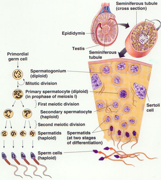
This diagram is to show the difference between the anterior and posterior pituitary - notice how the neurohypophysis is continuous with the hypothalamus and is made of neuro-secretory cells? While the adenohypophysis is almost like a separate organ dangling off the infundibulum - it is made of a different cell type and actually makes its own hormone products.


All three pictures here show the division between the adenohypophysis and neurohypophysis, What cells can you see in the last picture? You should be able to point out 5 kinds and know what they secrete. 

See the 3 different types of cells? Which ones are the acidophilic chromophils? Basophilic? Remember, cells that are acidophilic tend to stain brightly, or appear more 'warm' in color


-Thyroid/parathyroid: The parathyroid sits on the thyroid. Remember that the thyroid has minimal CT which is how you can distinguish it from the mammary. Where on the picture below would you find follicular cells? Parafollicular cells? What do they both secrete??

Thyroid Gland
Border between thyroid and parathyroid.
Parathyroid - You can see principal/chief cells, oxyphil cells and unilocular adipocytes in this shot. Can you point each of them out? Remember that chief cells are the smartest, look like brains (chromatin) and the oxyphil cells are more dense looking. 
-Pancreas - 
Where in this picture would you find beta cells? Alpha cells? What do they secrete? What secretes somatostatin? What does it do? Sorry for so many questions :) 
-Know the layers of the adrenal gland!! It's easy to identify the medulla, then if asked to point to a specific layer (zona glomerulosa, zona fasciculata, zona reticularis), point to the outside, middle or inside of the cortex, respectively. Know what each zone secretes (both the general class of hormones and a specific example, e.g. glucocorticoids and cortisol). The four pictures that follow are all of the same gland.

Human adrenal gland! It's important to keep the bigger picture in mind. 

Zoomed in... Can you name the three layers of the cortex and find the medulla? What doe the layers of the cortex secrete? The medulla?


Find the medulla..................
Trick question! There is no medulla in this shot, just doubled up cortex. See the pic below for an explanation.


Guys, please, please, PLEASE spend some quality time with the adrenal gland - not only is there a deceptively large amount of memorization associated with it but it will reappear in almost every bio class from here on out!!!! (Not even exaggerating!)

A mneumonic that helps me remember the zones of the adrenal cortex with the class and name of the hormone associated with them is:
Great Men Always Find Cute Girls Really Darn Attractive (eg. glomerulosa goes with the mineralcorticoid aldosterone, etc.)
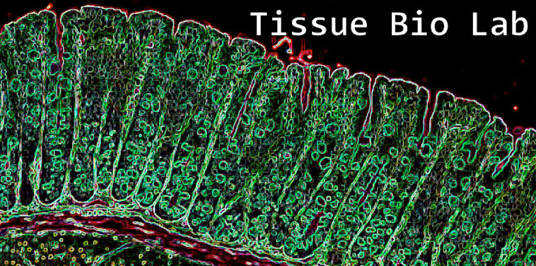

-Finally, the picture on p. 54 of your lab book is a cross section of a villus in the small intestine. You will find argentaffin cells all along the villus if you look. They are darker and wedge shaped. Know what they secrete!! Check out the picture below that is stained a bit differently than what you're used to seeing. The arrow is pointing to the enteroendocrine/argentaffin cell. Notice the difference between it and the goblet cells below it (goblet cells have no noticeable nucleus). 


Good luck! Let me know if you have any questions.

Endocrine Review Sheet
Key: Know the anatomical and histological names (including modifications) for the following bolded structures; assume that you will be required to find the structures indicated by * on your own slides.
***This list is not guaranteed to be exhaustive, and only includes terms from this unit. While we will not focus on quiz information from previous weeks, knowledge of previous material may be useful***
Focus on endocrine histology, but keep in mind all of the following terms. Know the endocrine secretions and functions as outlined in your manual.
Pituitary Gland
Anterior Pituitary (Adenohypophysis)
·       Pars distalis*
·       Chromophobes*
·       Basophilic chromophils*
·       Acidophilic chromophils*
·       Pars intermedia*
·       Pars tuberalis*
Posterior Pituitary (Neurohypophysis)
·       Herring bodies*
·       Pituicytes*


Thyroid
·       Follicular cells*
·       Parafollicular cells*
Parathyroid
·       Principal (chief) cells*
·       Oxyphil cells*
Pancreas
Know the 4 types of cells found in Islets of Langerhans, the general region where they are found, as well as their secretions
·       α-cells
·       β-cells
·       δ-cells
·       PP-cells
Stomach
·       G-cells
·       Parietal cell*
·       Chief cell*
Adrenal Gland
For each region of the adrenal gland, know the class of hormone that gets secreted as well and  a specific hormone example.
·       Zona glomerulosa*
·       Zona fasciculate*
·       Zona reticularis*
·       Medulla*
Enteroendocrine Cells
·       Argentaffin cells*