
Key things to remember:
-DCCTRA is by far the most common type of connective tissue in the organs that we are studying this week, if you can't remember what kind of connective tissue is present in a specific organ, you're best bet is to go with DCCTRA.
-Make sure you can identify the different fibers in areolar tissue (collagen, elastic) and also be able to ID fibroblasts and know what they secrete. Your manual gives a good explanation of this if you have any q's...
-Hyaline cartilage and elastic cartilage BOTH have chondrocytes, chondroblasts, and a perichondrium (layer on the very outside of the cartilage). The ONLY way to tell them apart is that elastic cartilage has elastic fibers (why it's DECTRA) which are only visible if the cartilage is stained with Orcein-Resorcein/Verheoff's stain. The elastic fibers are very dark because of the stain. Don't get confused though because hyaline cartilage can be stained darker (e.g. purple) too, but it will be a uniform darkness. See below for pics of how elastic fibers cause a non-uniform darkness from the stain.


Examples above are all Elastic cartilage. Notice the contrasting for the elastic fibers and the extracellular matrix.
 |
| Hyaline cartilage. Notice how the extracellular matrix (space outside of chondrocytes) is much smoother?? No elastic fibers...
-Fibrocartilage
has NO perichondrium, and most often does not have a noticeable
chondroblast layer (though it can...). It will have visible chondrocytes
if zoomed out, and a meshwork of irreg. fibers if zoomed in. Look in
your lab manual for a good example of both.
  |
Fibrocartilage. Notice the lack of a perichondrium/chondroblasts.
-Know where to find lamina propria!
In the intestinal tract (just deep to the epithelium layer before you
get to the submucosa) and in the oviduct (also just deep to the
epithelium layer).
-The
hepatic/glisson's capsule surrounding the liver is DCCTRA as are almost
all capsules. Students are often confused when they see a zoomed out
picture of the liver. It has many lobules and they are very noticeable
when zoomed out. Don't get confused if you see that!
 |
| See the lobules?? |
-Tendons have a
general waviness to them which is especially noticeable when zoomed out
further than the picture in the lab manual and they help to connect
muscle to bone. They can be other colors than pink as is shown in the
manual. They have NO horizontal striations which makes them look
different than muscle.


Both
photos above are of tendon. Compare them with the one below of skeletal
muscle. See the horizontal striations in the one below? That's one key
way to tell them apart.

-Ligaments
- Don't stress too much about them. Know that they connect bone to bone
and that they have elastic fibers (thus DECTRA instead of DCCTRA). What
stain would you use to make the elastic fibers visible?
-Know all the terms in the section about bones....
-Make
sure you can find endochondral bone formation before your quiz! Each
box has an endochondral bone slide, but it is often hard at first to
find where in the slide the endo. bone formation is occurring. These are
easy points as long as you can find it on your slide!
Remember: Real
People
Help
Cancerous
Rats
Some hints for each zone: zone of reserve (think reserved people stay by themselves) has mostly single/double cells that are fairly flat. Zone of proliferation (think, they are prolific
and having lots of kids...) shows mostly flat cells, but they are in columns with several others. Zone of hypertrophy, the number of cells in the columns don't change, but they are much bigger (more swollen looking). This layer is probably one of the best landmarks because it is usually the most visible. Zone of calcification is immediately after the zone of hypertrophy where there are no more visible cell membranes because the cells are becoming calcified. If asked to identify it, point immediately after the zone of hypertrophy or else you risk pointing to the zone of resorption. If asked to point to the zone of resorption, make sure you are not right next to the zone of hypertrophy.
-For
the developing tooth, pick a landmark! I like to use dentin as mine. It
is often the most recognizable and is always thick and colorful
(usually bright pink). Make sure to know that the ameloblasts secrete
enamel inward toward the tooth and that odontoblasts secrete the dentin
outward. Know what the ameloblasts and odontoblasts are histologically,
and make sure that if you are asked to point them out, that you are
pointing to the right layer! They are fairly obvious because they are
and look like simple columnar!


All
pics of developing teeth. Notice how the dentin layer is the most
obvious landmark in each?? (Top left=the lighter pink, smooth layer; top
right=bright pink thick layer; bottom=bright pink thick layer) I would
suggest identifying the dentin layer and then working out/in from there.
-For
the spleen, look for a thick capsule with lymphatic nodules as your
landmarks. Please do not think that white pulp is what looks white and
the red pulp is what's red!!! The white pulp is what is inside the lymph
nodules (see the pics below) and the red pulp is everything surrounding
the lymph nodules.

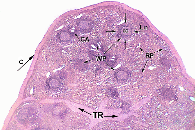
See the thick capsule? The circular parts are the lymph nodules.
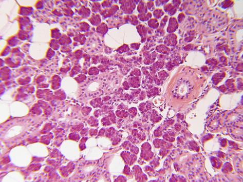
-The
internal elastic laminae is the squiggly dark line (stained with
orcein-resorcein) you see near the lumen of arteries (most obvious on
medium sized arteries) and the external elastic laminae is the
collection of dark squiggly lines a bit deeper once you get past the
smooth muscle. The tunica adventitia is immediately after the external
elastic laminae. When the artery is in diastole (relaxed), the squiggly
lines are also relaxed and they remain squiggly --> DECTIRA (squiggly
lines are irregularly arranged...). When the artery is in systole
(blood is higher pressure), the elastic laminae are stretched and are no
longer squiggly --> DECTRA (regularly arranged). YOU WILL NEVER see a
slide of an internal or external elastic lamina in systole (the animals
are no longer living, thus their hearts are no longer pumping... :) ).
-Finally,
make sure you can find some examples of adipose (unilocular adipocytes)
on your own slides. Some good places to check are on your parotid slide
or around vasculature.
Connective Tissue Key Terms
Key: Know the anatomical and histological names (including modifications) for the following bolded structures; assume that you will be required to find the structures indicated by * on your own slides.
***This
list is not guaranteed to be exhaustive, and only includes terms from
this unit. While we will not focus on quiz information from previous
weeks, knowledge of previous material may be useful***
Focus on connective tissue histology, but keep in mind all of the following terms.
Basic Connective Tissues
Aerolar Connective Tissue
· Aeolar connective tissue*
· Fibroblasts*
· Collagen fibers*
· Elastic fibers*
Hyaline Cartilage
· Hyaline cartilage*
· Chondroblast layer*
· Chondrocytes*
· Nest cells*
· Perichondrium*
Elastic Cartilage
· Elastic cartilage*
· Chondrocytes*
· Chondroblast layer*
· Perichondrium*
Fibrocartilage
· Fibrocartilage*
· Nest cells*
· Chondrocytes*
Digestive System
Intestinal Tract
· Lamina propria*
· Muscularis mucosa
· Submucosa*
Liver
· Glisson’s capsule*
Musculoskeletal System
Tendon
· Tendon*
· Collagen fibers*
· Fibroblast nuclei*
Ligament
· Ligament
· Elastic fibers
· Fibroblast nuclei
Bone
· Haversion canal*
· Osteocytes in lacunae*
· Canaliculi*
· Concentric lamellae*
· Interstitial lamellae*
Endochondral Bone Formation
· Zone of reserve*
· Zone of proliferation*
· Zone of hypertrophy*
· Zone of calcification*
· Zone of resorption*
· Perichondrium*
· Periostium*
Developing Tooth
· Dental pulp*
· Odontoblast layer*
· Predentin*
· Dentin*
· Enamel*
· Ameloblast layer*
Respiratory System
Trachea
· Chondroblast layer*
· Perichondrium*
· Tracheal epithelium
Dermis
· Dermis*
· Epidermal epithelium
Lymphatic Tissue
Spleen
· Splenic capsule*
· White pulp*
· Red pulp*
Lymph Node
· Reticular fibers
· Dense lymphatic tissue
Cardiovascular
Blood Vessels
· Internal elastic lamina (systole and diastole)*
· External elastic lamina (systole and diastole)*
· Tunica adventitia*
Miscellaneous
Adipose Tissue
· White adipose cells*
Oviduct
· Oviduct epithelium
. Lamina propria*


