Hey everyone!
I just wanted to post to make sure
everyone is up to speed on the midterm next week. The midterm will be
very similar to your other in-lab quizzes with a few minor exceptions.
First, (obviously) the midterm will cover everything that we have
covered so far this semester. Don't forget to go back and study the
development material (e.g. 24, 48, 72 hour chick slides). Also, the quiz
will have a few extra slide questions, which will make the "U-finds"
worth a half point each this week instead of a full point. To make it to
the ten points, we will also have two "We-Find" questions where we
point to a structure on a microscope in the back of the room, and you
have to identify the structure. The slide label will be covered and you
won't be able to change the zoom or move the slide. You can adjust the
focus if you need to :) Besides those small differences, the midterm
quiz will feel totally normal for you guys and will be great practice
for the final!
My biggest suggestion for doing well on
the midterm is identifying landmarks for each of the organs you have
learned (what makes each organ unique, ex: thymus vs. the liver, spleen
vs. lymph node, hyaline cartilage vs. fibrocartilage). The hardest thing
for students is often figuring out what organ the picture is showing.
If you have good landmarks for each organ, it will make this step a lot
easier. Once you figure out which organ you are in, attack the question.
If you are coming up blank on the question, it helps to think about
what structures you have learned for that specific organ and then go
from there. If you are having trouble finding a landmark on the slide
and identifying the organ, look for the most noticeable characteristics
(i.e. epithelium, glands, cartilage, etc.) and go through
process-of-elimination-style in your head which organs have those
characteristics (e.g. what structures have I learned that have
stratified squamous epithelium and glands in the submucosa??). Once you
get good at that, you are normally only a couple of steps away (at most)
from figuring out which organ you're in. If you are having trouble
coming up with good landmarks, feel free to contact us! My last
suggestion before I get off my soap box: try to look at as many pictures
of the structures as you can as you're studying. Looking at the same
picture over and over can only help you so much. The links on the right
are great for practice with this. Good luck! You guys are all going to
do great.

Tuesday, February 18, 2014
Friday, February 7, 2014
Blood & Lymph
I hope you all enjoyed looking at your own blood cells during class last week :) Good news, this week is one of the lightest of the semester. Take advantage of that and everyone get a 10 on the quiz! As always, feel free to submit a question if you have one!
Important things for this week:
-MEMORIZE the chart that was on the board with the info about relative amounts of each white blood cell, life span, and function. This will get you easy points on this week's quiz, the midterm, and the final. A good mnemonic to remember the different types of WBCs is "Never let monkeys eat bananas!!!"
-Make sure that you can identify each type of blood cell (all of the WBCs, erythrocytes, megakaryocytes, reticulocytes) in a picture, that you can find the most common cell types (erythrocytes, neutrophils, and lymphocytes) on your slides, and that you know what processes each cell type is involved in.
For instance - Neutrophils = acute inflammation - remember, they're the white stuff in pus
Eosinophils = parasitic invasion
Basophils = related to mast cells - very involved in allergies - remember, they secrete
histamine and heparin
Monocytes = they're all about eating
Lymphocytes = these are the T-cells and B-cells of immunity - they're involved with
your adaptive immune system
You can use your blood slide or one of the generic ones in your box.
- Also, which WBCs are granulocytes ("B-E-N")? agranulocytes (M-L)?
 |
| Cool EM of erythrocytes and leukocytes! Can you guess which are the WHITE blood cells?? :) |
 |
| What your blood slide will look like at about 4x mag. Zoom in on the purple spots (what are they stained with???) to pick out specific WBCs. |
 |
| What do you
see here? Here's a hint: the one at the middle-left is the MOST common
WBC (notice the multi-lobulated nucleus). The one on the right is the
second most common WBC (notice the big, dark nucleus with the halo-like
cytoplasm and no visible granules). What do you guys think the small
dark spots are? (Hint: They're the result of thrombopoiesis). |
 |
| Someone that has leukemia... SOO many lymphocytes. |
-Neutrophils are by far the most common and have he multilobulated nucleus (looks like several kidney beans connected by a small extension). Although most of your pictures in the manual only show 2 lobules, they can have several.
-Eosinophils can look similar to neutrophils, but are much brighter.
-Monocytes - remember the butt-print! (What it looks like when you're wet and sit on a dry chair and stand up).
-Basophils are dark and have lots of granules... usually so many that you can't see the nucleus.
 |
| Can you pick out all the different ones here? The two on the left are the same... Here's a hint, ID the following WBCs: neutrophil, basophil, lymphocyte, and eosinophil. |
-The key for the lymphatic system is knowing what makes each organ unique, because they all have the lymph nodules.
-Lymph nodules are characteristically darker around the outside edges and lighter on the inside.

-Besides the capsules and epithelium taught in this chapter, almost everything is histologically named dense lymphatic tissue.
-One common confusion: lymph nodes are larger than lymph nodules and house many lymph nodules within them.
-The center of the lymph nodule is the germinal center, and almost always will have lymphoblasts and monoblasts (except for in the spleen where they have t cells and b cells).
-What makes each organ unique:
- Lymph node: lymph nodules around the outside (cortex). The medulla does NOT have lymph nodules.
 |
| See how the lymph nodules are limited to the outside (cortex)?? |
- Ileum (Peyer's patches): Lymph nodules limited to the submucosal area. Look for the evaginations that are so characteristic of the small intestine. REMEMBER that peyer's patches are not found in the duodenum or jejunum, so if you see lymph nodules in the submucosa of the small intestine, it has to be the ileum.

- Appendix: trash in the lumen, epithelium (simple columnar w/striated...), lymph nodules in the submucosa.
 |
| Good shots of the appendix. Notice the invaginations as opposed to the evaginations in the small intestine. |

- Spleen: Can look similar to the other organs (lymph node, liver, etc.), BUT it is the only one that has lymph nodules spread throughout the entire organ (lymph node only has them around the outside, etc.). Remember that the spleen has unique cells in the germinal center (t & b cells).
 |
| See the nodules all over? Compare this to the lymph node. |
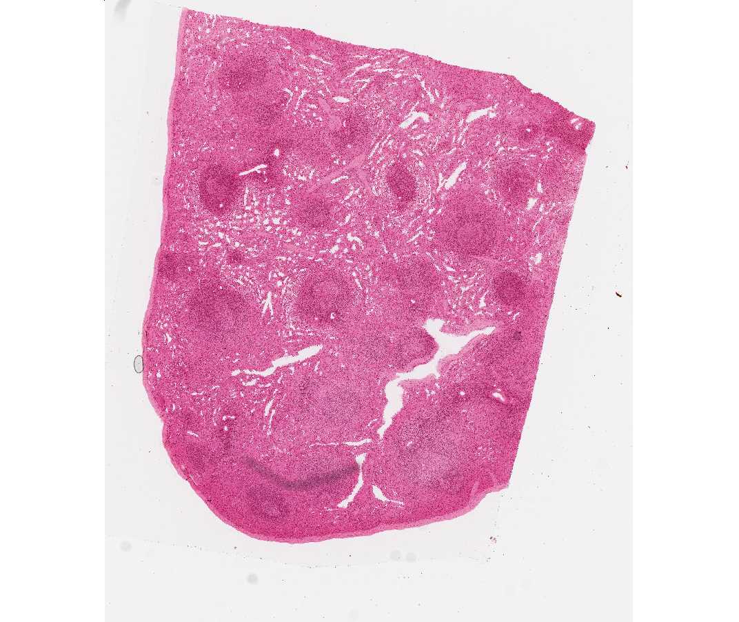
- Thymus: LOBES that have the characteristic darker outside with lighter inside. Hassall's corpuscles (noticeable cells at the center of the germinal center). Be careful, the thymus can look very similar to the liver, but is distinguishable because the liver does not have the lighter centers and darker outsides of the lobes. The liver can have vasculature running down the center of the lobes which can look like Hassall's corpuscle so keep that in mind for the midterm/final! Use the shading of the lobes as your guide. See how the bottom pictures look similar??? The one on the right is the liver, the left is the thymus. The point of differentiation is the shading of the lobes.
 |
| Hassall's Corpuscle - See how it looks different than vasculature? (no noticeable lumen, no RBCs |


- Tonsils: Epithelium=strat. squamous (why??), though not always visible. TONS of nodules, generally arranged in rows.

Good luck everyone!!
Terms List: (key: know the histological names for the structures with [h] after them; assume you must be able to find these structures on your own slides unless indicated by [x])
I also included links to digital microscope slides for several of the
structures. If the structure is harder to find, I set the link to
automatically zoom to the place with the structure. On those, it would
be wise to zoom out and make sure you can find it on your own. As
always, feel free to email me with questions!
***This list is not guaranteed to be exhaustive, and only
includes terms from this week. While
we will not directly quiz information from previous weeks, knowledge of
previous material will help a lot***
***Make sure you know everything on the chart that was on
the board about WBCs***
RBC (erythrocyte)
Erythropoiesis
Reticulocyte [x]
***A note about the WBC’s: You should be able to find a
neutrophil and lymphocyte on your own blood slide. It might be harder to find
the other three WBC’s. With that said, your blood smear slide in your box will
oftentimes have the others as well.
Neutrophil
Eosinophil
Basophil
Monocyte
Lymphocyte
Megakaryocyte [x]
Thrombocyte
Thrombopoiesis
Lymph node [h]
Medulla
Cortex
Lymph
nodule [h]
Germinal
center [h] – what cells are found there?
Ileum
Peyer’s
patches [h]
Germinal
center [h]
Appendix [h]
Lymph
nodule [h]
Germinal
center [h]
Spleen
White
pulp [h]
Red
pulp
Trabeculae
[h]
Thymus
Cortex
[h]
Medulla
Interlobular
septa [h]
Hassall’s
corpuscle
Tonsil
Lymph
nodule [h]
Germinal
center
Epithelium
[h]
Lymphatic vessel [h] [x]
Subscribe to:
Posts (Atom)