Tuesday, March 26, 2013

Digestive & Respiratory!

Hey everyone! This week's material isn't too bad. I think there are 3-4 new structures in the entire lesson. Take advantage of the lighter lesson to review, review, review. The final is just around the corner and it is not something that you'll want to cram for. In this post, I'm just going to highlight the new things we taught you guys this week and a few other important points. So here goes:

-You'll notice that this week we taught you guys the muscle layers surrounding the esophagus. It's pretty intuitive though since it is part of the GI tract: Inner circular, outer longitudinal.
Always keep in mind the relative anatomy of the structures we're learning about! Which is the trachea? Which is the esophagus? What would it look like if a pic of the esophagus included part of the trachea?
-Make sure you know how to distinguish between the different parts of the small intestine: duodenum (Brunner's Gland), Jejunum (nothing remarkable), Ileum (Peyer's Patches) [pictures below in that same order]


-One of the new things this week: MUSCULARIS MUCOSA. You can find it all over the digestive tract and it is a small muscle layer separating the mucosa and the submucosa.
5=muscularis mucosa, 6=submucosa, 4=lamina propria. Where in the GI are we?
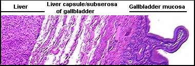
-Gallbladder - New this week too! Has evaginations and the simple columnar epithelium, but it doesn't have the striated border like the small intestine. Pretty unique looking. You can often see the liver in the slide as well (See the second pic for an example).


-Liver - We have obviously talked about this organ before, but we added hepatic sinusoids this week (histo=simple squamous). The sinusoids are just the white spaces between the hepatocytes (which are often binucleated). Von Kupffer cells are the phagocytic cells (mod. simple squamous) that sit in the sinusoids.
Lighter spaces=sinusoids, darker cells in sinusoids=Von Kupffer cells
-Know the trends for goblet cells: increase down GI tract (most concentrated in colon), decrease down respiratory tract (most concentrated in trachea).
-New this week for respiratory: Dust cells! These are super easy to find. Just look for an alveoli and then any renegade cell in the alveoli is going to be a dust cell.
What's labeled as an "alveolar macrophage" here is a dust cell which is mod. simple squamous.

I think that's it for this week. Let us know if you have any questions! Last week of new material this week!

Wednesday, March 13, 2013

Feeling hormonal???

If you are, I don't blame you. I know there is quite a bit of memorization for this week. Learn it! It will help you in almost every bio class you have here on out. This week the anatomical structures aren't too difficult, but make sure you know what hormone(s) they all secrete and the effect of those hormones.

Things to remember:
-The part of the pituitary that is connected to the stock is the neurohypophysis (posterior pituitary). The neurohypophysis doesn't produce any hormones, it simply stores them for secretion. While the adenohypophysis is usually darker/brighter (because of the acidophilic chromophils), color can sometimes be deceiving.


All three pictures here show the division between the adenohypophysis and neurohypophysis, What cells can you see in the last picture? You should be able to point out 5 kinds and know what they secrete. 
-Thyroid/parathyroid: The parathyroid sits on the thyroid. Remember that the thyroid has minimal CT which is how you can distinguish it from the mammary. Where on the picture below would you find follicular cells? Parafollicular cells? What do they both secrete??

Thyroid Gland
Border between thyroid and parathyroid.
Parathyroid - You can see principal/chief cells, oxyphil cells and unilocular adipocytes in this shot. Can you point each of them out? Remember that chief cells are the smartest, look like brains (chromatin) and the oxyphil cells are more dense looking. 
-Pancreas - 
Where in this picture would you find beta cells? Alpha cells? What do they secrete? What secretes somatostatin? What does it do? Sorry for so many questions :) 
-Know the layers of the adrenal gland!! It's easy to identify the medulla, then if asked to point to a specific layer (zona glomerulosa, zona fasciculata, zona reticularis), point to the outside, middle or inside of the cortex, respectively. Know what each zone secretes (both the general class of hormones and a specific example, e.g. glucocorticoids and cortisol). The four pictures that follow are all of the same gland.

Human adrenal gland! It's important to keep the bigger picture in mind. 

Zoomed in... Can you name the three layers of the cortex and find the medulla? What doe the layers of the cortex secrete? The medulla?


Find the medulla..................
Trick question! There is no medulla in this shot, just doubled up cortex. See the pic below for an explanation.


-Finally, the picture on p. 54 of your lab book is a cross section of a villus in the small intestine. You will find argentaffin cells all along the villus if you look. They are darker and wedge shaped. Know what they secrete!! Check out the picture below that is stained a bit differently than what you're used to seeing. The arrow is pointing to the enteroendocrine/argentaffin cell. Notice the difference between it and the goblet cells below it (goblet cells have no noticeable nucleus). 


Good luck! Let me know if you have any questions.

Friday, March 8, 2013

You've got some nerve.....

Sorry for the pun.... :)  You guys are all expert histologists now. Feels pretty good doesn't it?? If I were you, I would keep reviewing the midterm material over the next few weeks up until the final, because the new is definitely lighter than past lectures. One of the key things for this week is to recognize what nerve cells look like. Check out the pictures below. Notice that they all are fairly similar looking (as are most nerve cells)--central nucleus, large cytoplasm, etc.
The nerve cells are those running diagonally down the middle of the pic.

Other Key Things to Remember:
-The anatomical names of the nerve connective coverings are the same as muscle (epi-, peri-, and endoneurium), but the histological classifications are slightly different. Remember that the epineurium is the thickest covering that surrounds the entire nerve. Anything that is cutting through the middle of the nerve will be perineurium, and anything surrounding a single fiber/axon will be endoneurium.

-What stains nerve tissue??? (hint: Gold, _____, Bronze)

-Know what satellite cells are, where to find them (p. 46), and what they do.

-For the central nervous system, don't go off of the color of the tissue on the slides to determine what is white/grey matter because white matter can be the darker of the two... you think they would have figured that one out right??? :) Remember from your anatomy days that the brain is like an oreo (grey matter on the outside, white on the inside) and your spinal cord is like a hotdog (white on the outside and grey on the inside).

-Make sure you can ID the dorsal and ventral sides of the spinal cord. The easiest way for me to do that is to remember the pneumonic "Heading to the back dor (like door, stands for dorsal)." Let me explain... the dorsal horn is in the back, and it touches the edge of the spinal cord while the ventral horn does not. It looks like the DORsal horn is heading to the back door. Also, the dorsal horn is usually narrower. Whatever works for you!
See the dorsal horn heading to the back door???
-Make sure you memorize the layers of the cerebrum!!! And that you can name them in order... Don't worry too much about being able to distinguish between all the layers on the slide, they can be pretty ambiguous. Remember the PP sandwhich :) And that it's inner inner, outer outer with pyramids on the bottom and crumbling granules on top of them ( i.e. inner pyramidal, inner granular, outer pyramidal, outer granular).

-For the cerebellum, know the layers and how to ID them on a slide or on a U-Find. They're pretty logical though - white matter on the inside (remember... OREO!!), then comes the granular layer which looks.... very granular :) (always does...), and the outermost layer is the molecular layer (think: molecules are smaller than granules... can't really see molecules). In between the molecular and granular layers is the purkinje cell layer (looks like a row of nerve cells separating the two layers). Here are some pics for your viewing pleasure...

This picture should look familiar to all of those who are in physiology. Aren't those purkinje cells cool?!?! Which layer is more granular looking?

If you're having a hard time orienting yourself, look for the sulci (grooves of the brain).
- For the meninges, in between the lobes of the brain is a good place to point to for pia mater on U-Finds. Dura mater is often not present...

-Ependymal cells - where are they present??? (choroid plexus and central canal). Remember that they change histologically over time. I like the remember it this way: as you get old, you get short and bald (ciliated simple columnar --> sparsely ciliated simple cuboidal -->simple squamous).

-Auerbach's and Meissner's plexi both help regulating peristalsis. Auerbach's is found between the inner circular and outer longitudinal layers (think Auerbach sounds Austrian, kinda like Arnold Schwarzenegger, who has lots of muscles . . . so Auerbach's is gunna be between all those muscles). Zoom in and look for cells that fit the look of the ones at the beginning of this blog post. I prefer to look in the jejunum for these plexi, just because there aren't really any other structures there to confuse them with (no Brunner's or Peyer's patches). The Meissner's plexus is found at the very basal edge of the submucosa right before you get to the inner circular muscle layer (think Meissner Mucosa). Once again, zoom in and scan around until you find something that fits the profile of a nerve cell. 

Here's a little game of "Where's Waldo" for you. These pictures were both taken of that slide.
Meissner's (Base of sub-mucosa)

Auerbach's (lighter clustered structures toward the left-middle) - notice the smooth muscle on either sides??
Other Random Pics:

Meissner's

Auerbach's (notice the muscle layers on either side)


-For the retina: Be able to identify (slide and U-Find) all of the layers and name them in order. The inner/outer nuclear layers are highly nucleated. The ganglionic cell layer can also look nucleated, but will sometimes, but not always, be thinner than the nuclear layers; generally the ganglionic cell layer will have much larger nuclei. The plexiform layers look clear (like plexiglass). Remember that outer isn't referring to the outer part of the body, but the outer part of the slera (think the backside of your eyeball).

Notice how the ganglionic cell layer is thick here? (see E) However, the nuclei aren't as tightly packed, are larger, and have colored cytoplasm - fairly common characteristics for this layer


-Make sure you know what neurons are unipolar, bipolar, etc. Some people like to remember it using the following: one tongue --> unipolar neuron, two ears --> modified bipolar neuron, two nostrils-->bipolar neuron, two retinas --> bipolar neuron.

Good luck on the quiz!! As always, feel free to let us know if you have any questions.


Tuesday, February 19, 2013

Upcoming Midterm!

Hey everyone!

I just wanted to post to make sure everyone is up to speed on the midterm next week. The midterm will be very similar to your other in-lab quizzes with a few minor exceptions. First, (obviously) the midterm will cover everything that we have covered so far this semester. Don't forget to go back and study the development material (e.g. 24, 48, 72 hour chick slides). Also, the quiz will have a few extra slide questions, which will make the "U-finds" worth a half point each this week instead of a full point. To make it to the ten points, we will also have two "We-Find" questions where we point to a structure on a microscope in the back of the room, and you have to identify the structure. The slide label will be covered and you won't be able to change the zoom or move the slide. You can adjust the focus if you need to :) Besides those small differences, the midterm quiz will feel totally normal for you guys and will be great practice for the final!

My biggest suggestion for doing well on the midterm is identifying landmarks for each of the organs you have learned (what makes each organ unique, ex: thymus vs. the liver, spleen vs. lymph node, hyaline cartilage vs. fibrocartilage). The hardest thing for students is often figuring out what organ the picture is showing. If you have good landmarks for each organ, it will make this step a lot easier. Once you figure out which organ you are in, attack the question. If you are coming up blank on the question, it helps to think about what structures you have learned for that specific organ and then go from there. If you are having trouble finding a landmark on the slide and identifying the organ, look for the most noticeable characteristics (i.e. epithelium, glands, cartilage, etc.) and go through process-of-elimination-style in your head which organs have those characteristics (e.g. what structures have I learned that have stratified squamous epithelium and glands in the submucosa??). Once you get good at that, you are normally only a couple of steps away (at most) from figuring out which organ you're in. If you are having trouble coming up with good landmarks, feel free to contact us! My last suggestion before I get off my soap box: try to look at as many pictures of the structures as you can as you're studying. Looking at the same picture over and over can only help you so much. The links on the right are great for practice with this. Good luck! You guys are all going to do great.