
-You'll notice that this week we taught you guys the muscle layers surrounding the esophagus. It's pretty intuitive though since it is part of the GI tract: Inner circular, outer longitudinal.
 |
| Always keep in mind the relative anatomy of the structures we're learning about! Which is the trachea? Which is the esophagus? What would it look like if a pic of the esophagus included part of the trachea? |


-One of the new things this week: MUSCULARIS MUCOSA. You can find it all over the digestive tract and it is a small muscle layer separating the mucosa and the submucosa.
 |
| 5=muscularis mucosa, 6=submucosa, 4=lamina propria. Where in the GI are we? |

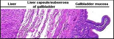
-Liver - We have obviously talked about this organ before, but we added hepatic sinusoids this week (histo=simple squamous). The sinusoids are just the white spaces between the hepatocytes (which are often binucleated). Von Kupffer cells are the phagocytic cells (mod. simple squamous) that sit in the sinusoids.
 |
| Lighter spaces=sinusoids, darker cells in sinusoids=Von Kupffer cells |
-New this week for respiratory: Dust cells! These are super easy to find. Just look for an alveoli and then any renegade cell in the alveoli is going to be a dust cell.
 |
| What's labeled as an "alveolar macrophage" here is a dust cell which is mod. simple squamous. |
I think that's it for this week. Let us know if you have any questions! Last week of new material this week!
No comments:
Post a Comment